


People and animals are attacked by these creatures, infections are easily transmitted to each other through infected products, water and dirty hands.The emergence of parasites in the body will help prevent precautionary precautions and maintenance regulations for personal hygiene, but these steps do not guarantee one hundred percent protection.Helminants who inhabit the human body can live in various organs, in contrast to the appearance, size, and degree of danger that people do.
Parasites in the human body
How many situations have been around when someone is walking around a doctor for years and unable to get rid of allergies, treat asthma, take sugar -guts and everything is worth it?Each of us has such contacts that spend a large number of treatments of various diseases, and does not get any results.
Only in rare cases, when the doctor is either very smart or very responsible, does he direct such patients to the usual analysis of stools and then ... then parasites are found in the human body, which causes dozens of diseases, and no one is fighting in this pathological treatment.
Contrary to the opinion, the worm is not necessarily "set" in the intestine and can be detected by the shallow analysis of the stool.Many parasites feel great in the lungs, heart, muscles, even in the brain and eyes.
The famous malaria, which has been defeated, has returned again and this is the most dangerous parasitic disease in the opinion of experts.Plasmodium malarial lives exclusively in the blood and not every doctor will be able to recognize the disease with sufficient confidence.
How parasites fall into the human body
They fall into the human body in different ways, often due to the use of water and infected foods.
The grape eggs remain viable for up to 6 months and through toys, rugs, underwear and pastel linen enter the body.Soldier eggs come to us in the vegetables and the non -washed fruits.Barbecue or homemade pig fat is a guarantee of 95% infection with trichinellosis.
Parasites penetrate us with insect bites, while bathing in fresh water reservoirs, through air, with dust, which is an egg carrier.
Salt fish, stroganin or caviar are the cause of infection with tape worms, reaching 12 meters and can live in your body under 25 years.The cases of infection have become more frequent in the parasites in the uterus.Dogs and cats, through their wet breathing, can remove parasite eggs at a distance of up to 5 meters.
You can be infected by dirty hands, not only their own, but also sellers, cooks, waiters, parasites traveling with money and public transport.High parasite egg concentrations are observed in products, such as: bacon, smoke sausage, ham, sausage, pork of any form, beef, chicken, goat, and even chicken eggs are infected with them.
Epidemiologists around the world are trying to fight this accident.In the United States, for example, for confirmation for helminthias, 1 pig car in each thousand is destroyed.This grows at a millions of losses, but if not, it's impossible.
There is no reliable way to eradicate meat, and the processing of ordinary larvae is not destroyed.You cannot guarantee yourself the purity of your food with welding or frying meat, a large number of parasite larvae still penetrates your body.
Where the organ can live in parasites
Helminthic parasites are divided into two categories that fit the place of activity in the donor body:
- Cramp: Worms living in various gastrointestinal parts.There are about 100 types of intestinal parasites, and for each bowel department there are several species of a dozen.The small intestine is ready to take ascaris, antelost, wide tape and other less common "relatives".The small intestine will "share the living room" with pinworms, dwarf chains and the rest.Medical literature describes the case when a person is infected with several types of parasites at the same time;
- Fabric:Worms are localized in organs, tissues, and even in the blood.Modern medicine successfully overcomes paragonimosis (lungs), cystycchosis (brain), echinococcosis (liver) and phylairiosis (lymph vessels).Some worms move through the body through the blood system and randomly attach to any organ.If many eggs have been introduced, then the whole body can be infected.
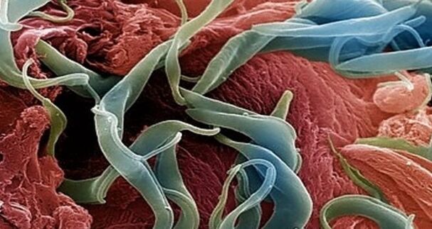
Snoes - How do they look in the human body (Worms photo)
Speaker- This is one of the most common human parasites, which is a circular worm (nematod).Often, infections with pinworms occur in children, but also in adults.
Pin is a white, small and round parasites.Individual female individuals have dimensions: 8-13 mm length, 0.5 mm thick, oval shape and straight tail, pointing at the end.
This feature of the female parasite tail describes her name - "cutter", from the word "sharp".Men's individuals are much smaller: the length is 2-5 mm, the thickness is 0.2 mm, the tail is bent, unlike the female pinworms.
Human invasion has the name of enterobiosis, and occurs primarily with non -compliance with the rules of personal hygiene (inadequate hand washing).Basically, pinworms live in small parts of the intestines and upper intestine, but in some cases they can also migrate to other organs and organs.

Women's helminth, who falls into the human body orally, and mates with nematod male representatives, migrated to the intestinal department, where she received the nutrients needed for the life and maturation of eggs from the waste of food.
After 4 weeks, the female cutter began to migrate in the rectum at a speed of 12 cm per hour, crawl out of the anus and put about 5000-15000 eggs in the perianal region, which after 4-6 hours completely cooked and ready for the next life cycle.
This process can be accompanied by itching, which encourages the infected person to brush the anus, and thus contribute to further spread of parasites, which fall into the food from the bottom of the nails, to the hands of others (especially for very close children in contact with each other and not always observe personal hygiene rules).
Infections with pinworms come from a person to a person, through dust with parasitic eggs, objects that are touched by the patient.Gutty eggs can also be transferred to foods with cockroaches and flies.
Also, the eggs remain on the linen, clothing, beds, which describe its fast distribution.Because the life cycle of the worm is so short, and the infection comes from a person to a person, it is difficult to get rid of parasites, because besides taking anthelmintic medication, the personal items of the patient are needed, and his insulation from other nematod media.
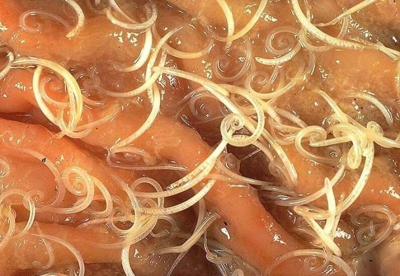
Soldiides - how they look in the human body (a picture of a worm)
Soldier-This is a large parasite shaped in the shape of a yellow spindle, reaching an adult 40 cm (female) and 15-25 cm (male).Without a suction cup or other setting device, ascaride can freely move towards the food mass.Eggs prescribed by female parasites are distinguished by impurities.
Acadosis infections occur if they swallow mature eggs together with water or vegetables -non -washable fruits where there is soil particles.After the eggs penetrate the intestines, the larvae cooks out of them.
Then, introducing it into the intestinal wall, they reached the liver according to the bloodstream, and from there they fell into the lungs.Through pulmonary alveoli, ascaride larvae through the respiratory tract penetrate the oral cavity again.
In the intestinal phase of their existence, ascarids are endowed with the ability to movement circles to penetrate narrow holes.These parasitic features often lead to the development of relatively serious complications (mechanical jaundice or pancreatitis)
After repeated swallowing, the parasite reaches the small intestine, where it develops into adults.The worm lived for 12 months, then died and stand out with stools.In the intestines of the owner can live both or a few hundred individuals.
Allergens secreted by ascarides can cause severe allergic reactions.Most adults can cause intestinal obstruction, and worms that have penetrated the respiratory tract sometimes cause drowning.

Vlasovsov - how they look in the human body
BlacovyvThey often live in the south, because these worm eggs love to heat.Most infections are observed in rural areas.Vlass -Head eggs live in the ground.
Invasia occurs by hand, contaminated particles, vegetables and non -washed fruits.As a result of the infection, the disease occurs - trichocephalosis.Vlashev parasites in the intestine.This worm causes anemia, as it consumes human blood, and severe abdominal pain.
For the diagnosis of trichcephalosis, the rectum and sigmoid are checked with special devices (rectoroscopy).Therefore, the accumulation of parasites in the intestine is found.Treatment of intrusion is long, as the wound is protected by a thick shell.
Parasite eggs stand out with stools, but they are very small, they are not visible even under a microscope.Only with very strong aggression, it is possible to detect eggs in stool analysis.In shape, they look like adjustable, have a brownish yellow color.
On 2 sides, the egg is a hole.What does a worm look like?It is very difficult to find them to live in bowel movements, as blazoles cannot live beyond the human body.Only with anthelmintic therapy can be seen in white worm stools.
Bacon hepatic - how it looks in the human body
The parasites that cause opisthorchiasis are flat worms that reach 7-20 mm long.
In the acute phase of helminthiasis, the patient has pain in the upper abdomen, rising body temperature, nausea, developing muscle aches, diarrhea, and possible skin rash.Parasitic larvae begins to grow after the eggs fall into fresh water (from swallowed snails).Then they penetrate the fish body (Mas fish, crucian carp, bream, roach).
Human infections occur when you eat infected fish meat that has not undergone adequate heat treatment.Hepatic bomb larvae from the small intestine penetrates the biliary tract and into the gall bladder, setting there with two suction cups.
Chronic courses opisthorchiasis are characterized by symptoms of hepatitis, inflammation of the bile duct, cholecystitis, impaired digestive tract, nerve disorders, weakness and increased fatigue.Parasites are a driver of the development of irreversible changes, and even after expulsion, patients do not pass the chronic inflammation process and functional disorders.
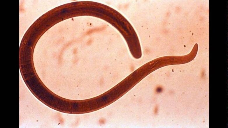
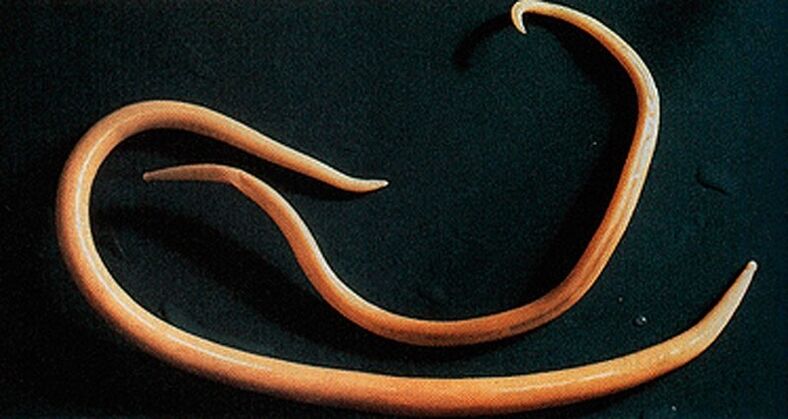
Trichinella - what appears in the human body (a picture of a worm)
The causative agent of trichinellosis is a small round helminth that reaches 2-5 mm long.Infections occur when the use of poor fried meat (pork, pigs, forest pigs).Penetrating the intestines, parasitic larvae within 3-4 days of cooking to sexually mature conditions.
The lifespan of the worm is 40 days, after which the parasite is dead.Driving the intestinal wall, the larvae penetrates the bloodstream and brought into all the organs of the human body, settling in the muscles.In this case, the respiratory and facial muscles, as well as the muscles of the limbs, are most often affected.
On the first day after the intrusion, the patient complained of stomach ache.
Then, after about 2 weeks, body temperature increased to 39-40 s, itchy rash appeared on the skin, muscle aches developed, and the face swollen.
During this time, in the case of large infections -there is a significant risk of death.After about a month, recovery occurs.The parasite is packed in the form of a circle, after which it dies within two years.
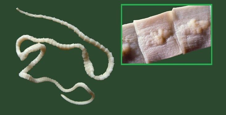
Wide tape - how it looks in the human body
This is one of the largest helminths that reaches 10-20 meters long.The disease caused by this parasite is called dipillobotriosis.The worm development cycle begins with freshwater fish or crustaceans.
Reaching the small intestine, the parasites are attached to the wall and for 20-25 days to grow into sexually mature individuals.
The larvae entered the human body, which is the owner of the extensive ribbon with the infected fish caviar or fillet.
Diaillobotriosis continues the background of the digestive tract disorders and B12 deficiency anemia.
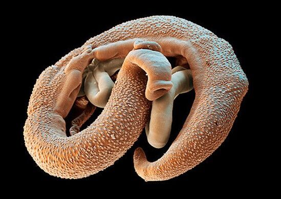
Echinococcus - how it looks in one's body
For this parasite, one is a mid -host.Parasitic worms in the human body in the form of finns.Echinococcus's final owner is a wolf, dog or cat.
Infections occur in a better way in contact with animals and environmental objects, with a handful of echinococcus eggs.After entering the intestines, oncospheres (six larvae) develop from them.From their intestines they penetrate the bloodstream and carry throughout the body.
The "favorite" places of worm parasitis are liver and light.Settling in these organs, the larva becomes finn (echinococcal cyst), which gradually increases in size, begins to destroy nearby fabrics.
Often, echinococcosis in the process of diagnosis is misunderstood as a benign or malignant original tumor.In addition to mechanical exposure (reducing organs and blood vessels), the rupture of the echinococcal cyst occurs sometimes.This condition can lead to toxic shock or the formation of various new cysts.
Alveococcus - how it looks in the human body
This parasite, which is considered to be a variety of echinococcus, is the cause of one of the most dangerous helminthiases (alveococcosis), which is similar to the severity of cirrhosis and liver cancer.Infection occurs with oncospheric penetration (eggs with cooked larvae) into the intestine.
These parasites, considered various echinococcus, are the cause of one of the most dangerous helminthiases (alveococcosis)
There, the embryo comes out of the egg and, introducing to the intestinal wall, penetrating the bloodstream.Further, with blood flow, parasites spread through all tissues and organs (most often localized in the liver).There the main developmental stage begins on the larvae (multi -chamber bubble, laurelocyst) formed).
Each space contains the parasite embryo head, which continues to grow gradually.Lavrocists are a very aggressive formation that is constantly growing due to increased bubbles, and also has the ability to sprout into the liver, such as cancer metastasis.
Nearby tissues are due to the operation of the affected blood vessels subject to necrotic changes.Opening a nearby structure, the alveokokk forms fibrous nodes with the entry of multi -chamber bubbles.This condition can last for several years, in relation to those who require compulsory surgical intervention.
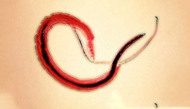
Schistosoma - How does the human body look like
Schistosoma - a blood, belongs to the trematodes, depending on the type that causes various schistosomosis.This is a separate helminth, reaching 4-20 millimeters long, 0.25 mm wide.The Schistosome body comes with a 2nd suction cup and stomach, they are located near each other.Schistos women are longer and thinner than men.There is a longitudinal groove on the man's body, so he holds a woman.Their eggs with a diameter of 0.1 mm, the shape of the oval, on the surface of one of the columns is a large spike.
The human worm schistosomes in the role of the final owner of choosing people, in their organisms they are parasites in small colon veins, abdominal cavities, uterus, bladder.Worms eat blood, partially absorb nutrients through cuticle.Eggs with Schistos are transported to the intestines and bladder, where they cook and stand out with stools or urine.In fresh water, larva - Miucidius, mid -host - mollusk out of eggs.In the mollusk body, Metacaria develops into 4-8 weeks.
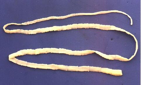
Pork Tinge - Like what human body looks like
Pork tape worms, such as bulls, on the body have 4 cups of suction, but in addition to this, the helminth body also comes with a double hook hook.Strogil reaches two to three meters long.Pork worms have three -doped ovaries, on each side of the uterus with 7 to 12 branches.The feature of this helminth is the ability of the segment to crawl out of the anus.After going out, their shells become dry and broken, so the helminth eggs enter the external environment.The middle master of the tape worm can be a pig and someone.
The main owner is the person.Intestinal parasites in humans include pig worms, helminth is located in the patient's intestines, where he lays his eggs.Infections occur when invasive meat is used.
Which doctor to contact with a worm to contact
If the infection with the worm is suspected, then it is necessary to consult Or parasitologist.You can contact an infectious disease specialist about infection with parasite or protozoa worms.
You can contact a helminthologist only if the infection is suspected exactly by the parasite worm (Pinworms, Ascarides, Plumings, OpisthorChiasis, etc.).
You can contact parasitologists in cases where protozoa infections are suspected - lamblia, toxoplasmes and amoebas.
In addition, if the parasite is localized not in the intestinal or stomach lumen, but in other organs (for example, the lungs, the liver), then you can contact the specialist involved in the diagnosis and treatment of this organ disease.For example, with opisthorchiasis, you can also contact a gastroenterologist or hepatologist, and with echinococcosis-to pulmonologists.